Monoclonal antibodies against biotinylable AP-tag / Avitag
Chemokine receptors undergo internalization and desensitization in response to ligand activation. Internalized receptors are either preferentially directed towards recycling pathways (e.g. CCR5) or sorted for proteasomal degradation (e.g. CXCR4). Scientists at the Georg-August-University Göttingen developed a method monoclonal antibody against a biotinylable peptid (Epitope-Tag) called AP-tag or Avi Tag, GLNDIFEAQKIEWHE. The 15-residue peptide served as a substrate mimic for biotin ligase (BirA), which usually recognizes the much larger protein domain. Anti-AP antibodies are useful tools in the analysis / trafficking of AP-tag fusion proteins, e.g. quantification of receptor-internalization from the cell surface into the cell. Binding of EF10 antibodies to the target protein is not affected by biotinylation of the acceptor sequence. Using these antibodies an alternative method was developed to analyse receptor internalization and recycling which allows a more detailed analysis of receptor trafficking compared to classical antibody-based detection methods. This biotin-based detection system may be generally applicable to the analysis of transmembrane protein trafficking.
Characteristics
| Clone: | EF10 |
| Specifity: | AP-tag, GLNDIFEAQKIEWHE |
| Isotype: | IgG1/k |
| Immunogen: | GLNDIFEAQKIEWHEC-KLH |
| Source (Species): | BALB/c mouse |
| Fusion Partner: | X63Ag8.653 myeloma cells |
| Species Cross Reactivity: | not tested |
| Concentration: | 35µg/ml |
| Applications: | Immunofluorescence staining, ELISA, Biological Function (Endocytosis Assay), Flow Cytometry |
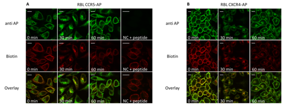
Double immunoflourescence (anti-AP/streptavidin) of CXCR4-/CCR5-expressing cells during ligand-induced internalization (30’) and recycling (60’).
Prior to stimulation and staining CXCR4-AP (right) and CCR5-AP (left) expressing RBL cells were seeded on glass cover slips. Cells were enzymatically biotinylated and stimulated with the corresponding ligand (125 nM CCL5, CXCL12; 30’/37°C). Recycling was induced by acid wash and transfer into ligand-free medium containing receptor antagonists (30 µM AMD 3100; 3 µM TAK779). Cells were fixed with 3% PFA (15’/37°C) and permeabilized with 0.1% saponin (15’/37°C). 10 µg/ml anti-AP antibody (anti AP; with or without preincubation with 2mg /ml of the AP-peptide) and 2 µg/ml streptavidin-Alexa647 (Biotin) was used for staining (60’/on ice/dark). Samples were fixed with mounting medium and analyzed by confocal laser scanning microscopy. Scale bar 10 µM.
References
Liebick M, Schläger C, Oppermann M (2016) Analysis of Chemokine Receptor Trafficking by Site-Specific Biotinylation. PLOSOne 11(6):e0157502.
Developmental Status
We are looking for companies, who are interested in licensing these antibodies for selling them to industrial and scientific institutions or for developing advanced diagnostic tests and therapeutic solutions.
Kontakt
Dipl.-Biol. Christiane Bolli
Licensing Manager Research Materials
E-Mail: Diese E-Mail-Adresse ist vor Spambots geschützt! Zur Anzeige muss JavaScript eingeschaltet sein!
Tel.: +49 551 30724 156
Referenz: BioM-1971-UMG
Tags: Antikörper